A Real Time-Saver speeds up your Research!
Multiple Application Scenarios -
- Cell Count Analysis: Suitable for counting cells or particles with a diameter of 4-60μm, such as most cell lines, stem cells, primary cells, etc.
- Cell Viability Analysis: When testing cell viability in trypan blue mode, after staining, live cells will be circled in green, and dead cells will be circled in red.
- Cell Size Analysis: In cell research, cell size is a key feature, which is often measured in cell transfection, drug testing and cell viability.
After counting, it will count the average diameter of the cells in the sample and automatically generate a histogram.
Cell Fluorescence Analysis -
- Detect Cell Transfection Efficiency (GFP, RFP): When transfecting cells with the target gene, the transfection efficiency needs to be evaluated. In general, the transfected plasmid has the sequence encoding the fluorescent protein as the reporter gene, so the successfully transfected cell should be positive for the fluorescent protein. In addition, using a fluorescence microscope or a flow cytometer is a commonly used method for transfection efficiency analysis in laboratories, but both have the disadvantages of complicated operation and high cost. Using the fluorescence analysis function of our system, equipped with the corresponding fluorescence module, can realize the analysis of the expression of GFP, RFP, and other fluorescent proteins, and automatically count them, so as to quickly, simply, and efficiently obtain the positive rate of target gene expression.
- Cell Fluorescence Analysis: Cell Viability Identification (AO/PI Dual Fluorescence Method). In addition to trypan blue staining, AO/PI dual fluorescence staining has become popular as a more accurate live cell counting method and is widely used for primary cell counting.
Highly Accurate
- High-definition imaging system & independently developed recognition algorithms technology enables it to count and analyze stem cells, primary cells and cell lines accurately.
- User-defined function. Users can self-define and preset thresholds for cell size, brightness, and roundness to reduce counting errors.
- Parameters can be modified at any time to obtain the best cell imaging & more accurate counting results.
- Primary mouse macrophages and HEK293 were counted with a hemocytometer and automated counters. The standard deviation of our system was smaller and the CV value was less than 5%>
Fast
- In bright field mode, a count can be completed in 9 seconds, while manual counting usually takes up to 5 minutes.
- Fluorescence analysis operation is easy to use.
- Built-in dilution calculator can give a dilution solution quickly based on the existed counting result, facilitate the experiments and save a lot time for operators.
- If each operator counts 10 cell samples (5pcs) per day, then 24.25 hours can be saved each month.
Convenient
- Multiple data display and storage, including cell images, diameter distribution histograms, count results, live and dead cells percentage.
- Convenient for storage and analysis, PNG, JEPG, CSV, PDF, etc.
- 1,000 historical counts can be stored in the device or exported via USB.
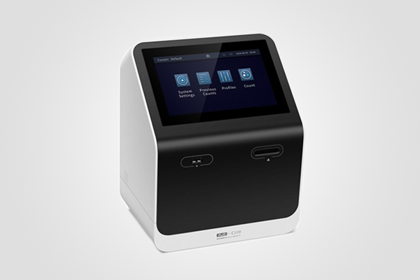
- 45° elevation angle, ergonomic design, reduced visual fatigue and cervical pressure.
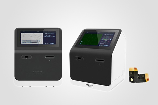
- 7-inch high-definition touch screen with friendly interaction and comfortable operation.